What each project asked, what I did, and what we found.
Most of my research so far has been about one inherited condition, oculocutaneous albinism type 1, from two directions: first measuring what it does to vision, then testing a gene therapy meant to treat it. This page explains each project, what I contributed, and what we found, so you don't need to read the papers to follow the work.
Background: oculocutaneous albinism type 1
Oculocutaneous albinism type 1 (OCA1) is caused by mutations in both copies of the gene for tyrosinase, the enzyme that starts melanin production. It affects roughly 1 in 36,000 to 40,000 people. Without tyrosinase, the skin, hair and eyes make little or no pigment.
In the eye, pigment normally sits in the iris, the retinal pigment epithelium (RPE, the cell layer just behind the light-sensing photoreceptors) and the choroid (the blood-vessel layer behind that). It absorbs stray light and cuts glare. Without it, people with OCA1 have painful light sensitivity (photophobia), glare and low vision. Missing pigment also disrupts how the retina develops. No approved therapy restores eye pigment.
Both studies below use the same OCA1 mouse model (B6(Cg)-Tyrc-2J/J), which carries a loss-of-function tyrosinase mutation and makes no pigment. Mice have no fovea, but the model shares the hypopigmentation and retinal developmental defects seen in patients, which makes it a widely used model for testing OCA1 therapies.
Measuring what OCA1 mice can see
Kriebel WG*, Larimer-Picciani AM*, Nukala M, Sahel J-A, Byrne LC. Quantifying functional vision in a mouse model of oculocutaneous albinism type 1. Scientific Reports 16, 14563 (2026).
Studies had shown that OCA1 mice are light-sensitive and have poor depth perception, but nobody had measured higher-level vision in this model, such as recognizing objects or resolving spatial detail. Without that baseline, there is no functional test to show whether a therapy actually improves vision. We asked which visually guided behaviors are impaired in OCA1 mice, and whether we could measure them reliably enough to serve as endpoints for future treatments.
What I did
I co-conceived the study with Alessandra Larimer-Picciani and Leah Byrne.
I developed the behavioral methods with Alessandra Larimer-Picciani.
I ran all of the behavioral experiments.
I analyzed the data with Mahija Nukala and Alessandra Larimer-Picciani.
I co-wrote the original draft and the revisions, and I presented the work at the University of Pittsburgh Ophthalmology Vision Research Day in 2024.
Approach
Three assays, three aspects of vision. We compared OCA1 mice with normally pigmented wild-type mice (C57BL/6J). In two assays we also tested rd1 mice, whose photoreceptors degenerate early in life, as a reference for little or no vision.
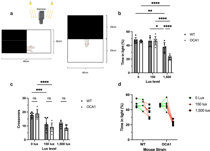
Light–dark box (photophobia). Mice moved freely between a dark compartment and a lit one at 0, 150 and 1,500 lux. We measured time spent in the light and the number of crossings into it (6 wild-type and 7 OCA1 adult males).
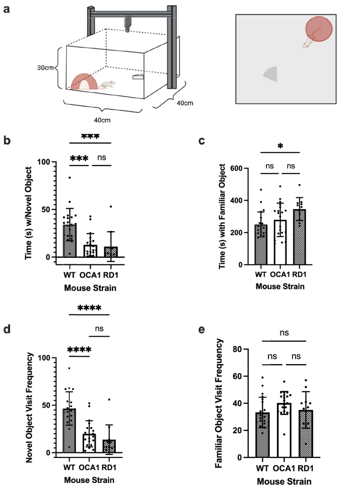
Novel object test (object perception). After the mice got used to a familiar object, we added a new one under moderate light (150 lux, below the level that triggers light aversion). We measured time spent near each object and visits to it (20 wild-type, 20 OCA1 and 11 rd1 mice).
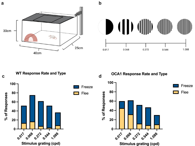
Looming stimulus with spatial gratings (spatial acuity). An overhead screen showed an expanding disc, the kind of shape that mimics an approaching predator. It was overlaid with striped patterns of five spatial frequencies, from 0.017 to 1.088 cycles per degree. We scored each response as freeze or flee and timed how long it took (20 mice per group).
Behavior was video-tracked and scored in EthoVision XT, looming stimuli were generated in PsychoPy, and freeze and flee responses were classified with objective velocity thresholds.
What we found
Light aversion appears only in bright light. At 0 and 150 lux, both groups split their time evenly (about 46–47% in the light). At 1,500 lux, OCA1 mice spent 22.6% of the trial in the light compared with 37% for wild-type mice (p < 0.0001), about 45 seconds less out of five minutes. Crossings into the light did not drop, so the mice still explored but chose not to stay.
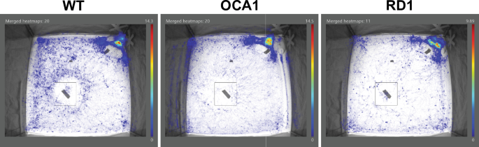
Object recognition is impaired even in moderate light. Wild-type mice spent 34.1 seconds near the novel object; OCA1 mice spent 12.8 seconds, statistically indistinguishable from blind rd1 mice (11 seconds). Wild-type mice approached the novel object 46.6 times on average, 2.3 times as often as OCA1 mice (19.9 visits). Heat maps showed wild-type mice circling the new object while OCA1 and rd1 mice rarely engaged with it.
Spatial vision is reduced, and responses are slower and different. OCA1 mice responded to looming stimuli 53% less often overall than wild-type mice (p = 0.048), although no single stripe pattern differed significantly on its own. Both groups were most responsive to the same stripe frequency (0.068 cycles per degree). OCA1 mice tended to flee rather than freeze (64 flee and 86 freeze responses, against 29 and 137 for wild-type), and they responded more slowly, for example 2.6 seconds versus 1.4 seconds at the coarsest pattern. rd1 mice did not respond at all.
Figure 1. Light–dark box. OCA1 mice avoid the lit compartment only at 1,500 lux, while their number of crossings stays the same.
Figure 1 from Kriebel, Larimer-Picciani et al., Scientific Reports (2026), reproduced unchanged under CC BY-NC-ND 4.0.Figure 2. Novel object test at 150 lux. OCA1 mice spend less time with the new object and visit it less often than wild-type mice, much like blind rd1 mice.
Figure 2 from Kriebel, Larimer-Picciani et al., Scientific Reports (2026), reproduced unchanged under CC BY-NC-ND 4.0.Figure 3. Where each group spent its time. Wild-type mice cluster around the new object (outlined); OCA1 and rd1 mice largely avoid it.
Figure 3 from Kriebel, Larimer-Picciani et al., Scientific Reports (2026), reproduced unchanged under CC BY-NC-ND 4.0.Figure 4. Looming stimulus with spatial gratings. The arena, the five stripe frequencies, and the rate and type of response for wild-type and OCA1 mice.
Figure 4 from Kriebel, Larimer-Picciani et al., Scientific Reports (2026), reproduced unchanged under CC BY-NC-ND 4.0.
Why it matters
This study gives the OCA1 mouse a behavioral profile that goes beyond light sensitivity. It shows deficits in object recognition and spatial processing even in ordinary light, and it provides quantifiable tests for whether a therapy improves vision. The light–dark assay from this work became the functional test in the gene therapy study below. The underlying causes still need work; the paper points to photoreceptor and retinal ganglion cell changes and to processing in the brain’s visual centers as next questions.
Restoring eye pigment with AAV gene therapy
Larimer-Picciani AM, Jacob LB, Sullinger KJ, Kriebel WG, Sahel J-A, Byrne LC. Systemic, RPE-directed AAV-tyrosinase therapy restores ocular pigmentation in an OCA1 mouse model. Molecular Therapy Advances 34, 201861 (2026).
My role: Co-author; I ran the behavioral experiments
One working copy of the tyrosinase gene is enough to make pigment, and the eye is easy to reach with adeno-associated virus (AAV), a harmless virus used to deliver genes, which makes OCA1 a good candidate for gene replacement. Earlier work showed AAV-tyrosinase could partly re-pigment the eye, but not how best to deliver it. This study asked which delivery route and which promoter (the genetic switch that controls where the gene is expressed) restore the most pigment in the iris, RPE and choroid with the least expression elsewhere, and whether the restored pigment changes how the mice respond to light.
What I did
I performed the behavioral experiments. I ran the light–dark photophobia assay comparing mice treated with the lead therapy against untreated littermates, using the same assay as the vision study above.
Alessandra Larimer-Picciani led the study and ran the other experiments, with TEM by Lucas Jacob and immunohistochemistry by Kacey Sullinger.
Approach
Two constructs. The mouse tyrosinase gene was paired with either a ubiquitous promoter (CAG, active in most cells) or an RPE-directed promoter (VMD2).
Three delivery routes. Intravenous injection one day after birth, before the blood–retina barrier closes. Subretinal injection (under the retina) at one month of age. Intravitreal injection (into the gel of the eye) at one month of age, using an engineered capsid designed to cross the retina’s inner surface.
Measuring pigment and safety. Pigment was assessed by fundus photography, whole cleared eyes, the pigmented area of flat-mounted RPE and choroid, and electron microscopy of melanosomes (the organelles that make and store melanin). Tyrosinase expression was measured in the eye, liver and brain by RT-qPCR.
Function. The lead therapy was tested with the light–dark box at 0, 150 and 1,500 lux.
What we found
Delivery route mattered most. Intravenous delivery pigmented the back of the eye most widely: 56% (CAG) and 65% (VMD2) of the RPE and choroid surface. Subretinal injection pigmented about 29%, concentrated around the injection site. Intravitreal injection left the back of the eye essentially unpigmented, although the CAG version densely pigmented the iris.
The RPE-directed promoter matched the ubiquitous one with far less off-target expression. VMD2 restored as much pigment as CAG while raising tyrosinase expression much less outside the eye: 34-fold versus 255-fold in the liver, and 2-fold versus 36-fold in the brain region containing the substantia nigra, where stray tyrosinase can be harmful.
Treated eyes made mature pigment, and it lasted. Electron microscopy showed mature, pigmented melanosomes in the RPE and choroid of treated mice, which untreated OCA1 mice lack entirely, though still fewer than in normally pigmented mice. Pigment was maintained five or more months after treatment.
In a small pilot, treated mice did not avoid bright light (my experiment). In a pilot of 3 treated and 4 untreated mice, untreated mice spent less time in the light as it got brighter (56.9%, 43.9% and 22.1% at 0, 150 and 1,500 lux). Treated mice generally showed no preference at any brightness (39.1%, 42.4% and 45.0%). The interaction between treatment and brightness was significant (p = 0.0156), but with this few animals no single light level reached significance on its own.
Graphical abstract. The study design and the lead strategy, intravenous RPE-directed AAV-tyrosinase.
Graphical abstract from Larimer-Picciani et al., Molecular Therapy Advances (2026), reproduced unchanged under CC BY-NC-ND 4.0.Figure 3. Whole cleared eyes, front and back. Intravenous delivery pigments the back of the eye widely; subretinal delivery pigments around the injection site; untreated OCA1 eyes stay clear.
Figure 3 from Larimer-Picciani et al., Molecular Therapy Advances (2026), reproduced unchanged under CC BY-NC-ND 4.0.Figure 5. Pigmented area of the RPE and choroid by route and construct, and tyrosinase expression in the front and back of the eye.
Figure 5 from Larimer-Picciani et al., Molecular Therapy Advances (2026), reproduced unchanged under CC BY-NC-ND 4.0.Figure 7. The light–dark experiment I ran. Untreated mice avoid the light at 1,500 lux; mice given intravenous AAV9.2yf-VMD2-Tyr show no clear preference.
Figure 7 from Larimer-Picciani et al., Molecular Therapy Advances (2026), reproduced unchanged under CC BY-NC-ND 4.0.
Why it matters
This is the first systematic comparison of delivery route and promoter for restoring eye pigment in an OCA1 model. It identifies RPE-directed AAV-tyrosinase as achieving widespread pigment with lower off-target expression, plus preliminary evidence that restored pigment changes light-avoidance behavior. The paper is direct about the limits. Newborn intravenous delivery isn’t an option in people, because the human blood–retina barrier matures before birth, and it carries systemic risk. So the next steps point to local delivery: subretinal injection, which is already used clinically, and suprachoroidal injection.
Other work in the Byrne Lab
From 2020 to 2025 I also worked on projects in the Byrne Lab that weren’t published as their own papers. Here I describe what I did on each.
Improving repigmentation in OCA1 mice
Brackenridge Summer Research Fellowship, 2021
I evaluated transcription factors and injection methods aimed at making AAV-mediated repigmentation more efficient in OCA1 mice. The University of Pittsburgh’s competitive Brackenridge Fellowship supported it as an independent undergraduate project. The work contributed to improving repigmentation efficiency, part of the lab’s broader effort to restore eye pigment described above.
Single-cell transcriptomics of tyrosinase-negative retina
I built a Python and R pipeline to analyze single-cell RNA sequencing (scRNA-seq) data, comparing gene expression in retinal cells from tyrosinase-negative and wild-type mice. The aim was to see which retinal cell types change when pigment is missing.
Computational prioritization of AAV capsid variants
I applied computational modeling and molecular docking workflows to rank novel AAV capsid variants (capsids are the protein shells that determine which cells a vector reaches). The rankings helped decide which candidates to carry forward for targeted gene delivery.
Producing research-grade AAV
I produced AAV vectors for preclinical studies. That covered HEK293 cell culture and transfection under BSL-2 conditions, large-scale purification by ultracentrifugation and ÄKTA FPLC with affinity and ion-exchange chromatography, and sterile formulation.